Home | Evidence | Anatomy | Skull Base
SKULL BASE [CRANIOCERVICAL] ANATOMY

INTRODUCTION
In order to understand the types
of problems and symptoms one might experience as a result of an atlas
to skull subluxation it's necessary to have a close up look at
the junction between the skull and the first cervical vertebra or atlas
[craniocervical junction]. At first glance it's clear that there
is not a lot of room at the skull base junction, in fact it's
much like a road map with nerves, blood vessels and ligaments crisscrossing
the area. Thus it is not hard to imagine that misalignments between
skull and atlas may have a significant negative affect on the critical
neurological and vascular structures whose pathways to and from the
brain pass through and around this area.
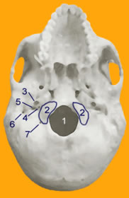 Figure 1: The HUMAN SKULL
BASE Figure 1: The HUMAN SKULL
BASE
Opposite is a depiction
of the skull base and anatomy of interest. This is the inferior
view or view from the bottom. The following structures or cavities
(foramen - holes) can be identified. These foramen and therefore
the neurological and vascular structures, which pass through them,
are in close proximity with the atlas vertebra and muscles and
ligaments attached to and surrounding the atlas.
- Foramen magnum
- this is the exit hole from the brain for the spinal
cord and entry to the brain for the vertebral arteries from
the cervical spine.
- Occipital condyles -
these kidney shaped structures articulate or connect to the articular
facets of the atlas, as shown in my section entitled "Anatomy
of the Atlas Subluxation" - reference 'superior
articular - occipital condylar surface'. To use an analogy,
these occipital condyles 'balance' on the atlas much
like one might place one's feet in horse stirrups. The stirrups
being the atlas facets and one's feet being the occipital
condyles.
- Carotid canal - this cavity is where
the internal carotid artery passes through into the skull in the
petrous temporal bone. The temporal bone is that which 'houses'
your hearing mechanism - internal ear structures. The carotid
artery runs through this bone very close to the pharyngotympanic
(auditory or Eustachian) tube. It may be possible for altered flow
of blood in the carotid artery to be detected by your inner ear
mechanism, and what affect does this have on your brain. Fernandez
Noda et al, in their paper 'Neck and brain transitory vascular
compression causing neurological complications', J CARDIOVASC
SURG 1996; 37 (SUPPL. 1 TO No. 6): 155-66, suggest "compression
of arteries including the carotid at the level of the cervical atlas
results in "faulty irrigation of blood supply and oxygen of
the cerebellum and basal ganglia of the brain. Among the effects
are: a decrease in the secretion of dopamine at the level of the
putamen, which produces the symptoms of Parkinson's disease"
etc.
- Hypoglossal canal -
this is where the Hypoglossal or cranial nerve XII exits the skull
after making its way from the medulla (lower portion of the brain
stem). The hypoglossal is a motor nerve and provides motor control
to the tongue muscles.
- Jugular canal - this canal contains
three major cranial nerves and a vein, these being;
- The glossopharyngeal
or cranial nerve IX, which is both a motor and sensory nerve.
It is responsible for visceral motor & sensation and taste.
It innervates the posterior tongue, walls of the pharynx and
the middle ear. The stylopharyngeus muscle is also innervated
by the glossopharyngeal. This is an interesting connection as
the stylopharyngeus muscles open and close the pharyngotympanic
tube by affecting the recoil of the cartilage around the tube.
Maybe here lies a connection to hearing disorders?
- The vagus or cranial
nerve X, which is the largest cranial nerve. Vagus is Latin
for 'wanderer' and this is because the vagus, being
a motor and sensory visceral nerve has major responsibilities
throughout the body and hence 'wanders' a long way.
This nerve sends motor control and receives sensory feedback
from the bile and gall ducts attached to the liver, the pancreas,
spleen, stomach, intestines, lungs, heart, and bronchial structures.
The vagus also provides sensation to the external acoustic meatus,
which is the ear canal.
- The spinal accessory
or cranial nerve XI is a motor nerve, which controls the neck
& shoulder muscles, such as the trapezius and sternocleidomastoid
but also joins with the vagus to innervate the muscles of the
larynx and pharynx.
- The jugular vein.
- Stylomastoid canal -
the facial nerve or cranial nerve VII passes through this foramen
on its way to innervate the muscles of facial expression. It also
innervates the stapedius muscle of the middle ear, which acts to
dampen the response of the ossicles of the ear to loud noises. There
are other functions of the facial nerve, which are listed in most
medical textbooks associated with this subject.
- Condylar canal
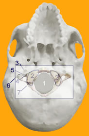 Figure
2: SKULL BASE with ATLAS OVERLAY Figure
2: SKULL BASE with ATLAS OVERLAY
In this figure I have overlaid the
atlas to show its position relative to the foramen and hence critical
structures leaving or entering the skull. In a non-subluxated position
the foramen are quite close and remember that there are many muscles
and ligaments between the atlas and the skull, which the neurological
and vascular structures must pass through. Thus when a subluxation,
even a minor one, exists at this level there may be quite dire consequences
for the individual. It is quite possible for the ligaments 'straining'
to maintain stability of the upper cervical spine and hold the head
perpendicular, to place compressive or traction forces on the cranial
nerves and blood vessels in and around these ligaments or muscles. The
result can be attenuated nervous system signals and/or attenuated blood
flow. What would be the result of the vagus not firing at full potential?
It is not hard to envisage organs, which do not function correctly.
And what would be the result of a reduction in blood flow to the brain?
We know that in catastrophic occlusions of arterial flow stroke can
be a result. What would the result be even in minor reductions in flow?
I suggest the result could be myriad neurological and/or other symptoms,
which, apart from the complaints being made by the patient, go undetected
by the diagnostic processes available to your average MD or specialist.
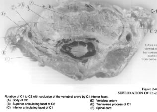 Figure 3: C1-C2 SUBLUXATION
and OCCLUSION of VERTEBRAL ARTERY Figure 3: C1-C2 SUBLUXATION
and OCCLUSION of VERTEBRAL ARTERY
Opposite is an axial section (or cut)
as seen from below of a subluxation of C1 (atlas) to C2 (axis). It is
noted by Ruch, "it is apparent that C1 is rotated relative to
C2 and the inferior articulating facet of C1 on the left is contacting
and compromising the vertebral artery." Given that the vertebral
artery provides blood supply to the cervical spinal cord and posterior
portions of the brain, e.g. cerebellum, surely this kind of subluxation,
which I suggest is quite common, would have the potential to result
in adverse and even dire consequences for an individual?
SUMMARY
The base of the skull, that
is, the junction between the skull and the upper cervical spine, is
packed with very critical neurological and vascular structures. These
structures wind their way to and from the brain via foramen or holes
in the base of the skull and pass through ligaments and muscles responsible
for maintaining the head atop of the cervical spine (neck). When a person
receives a blow or bump to the head, the result can be a shift of the
skull on the first cervical vertebra (atlas or C1). This shift causes
the ligaments and muscles holding the head perpendicular to the spine
to go into spasm, which can and does result in compression and/or traction
of the vital nerves and blood vessels passing through them on their
way to organs and other parts of the body. Long-term affects of this
on the body, amongst other things are biomechanical changes to the spine,
viewable postural changes, atrophy of key muscles e.g. trapezius &
sternocleidomastoid and dysfunction in organs. Tension can be placed
on the lower part of the brainstem or upper part of the spinal cord,
which of itself alone is a cause for concern. Anyone who has sustained
a subluxation of this kind, and I'm such a person, will attest
to the multiple and distressing symptoms which seem to abound. Those
like me that have run the gauntlet of modern conventional medicine in
search of answers will also attest to a lack of understanding of this
injury. I was lucky enough to stumble upon 'specific' upper
cervical chiropractic as a solution to this problem, but not before I was subjected
to unnecessary surgery, in the "off chance" that it may
relieve me of my symptoms, which it was being suggested were all in
my mind. Have a look at my symptoms at the time. One would have to conclude
some kind of cranial nerve or vertebral artery involvement. I don't
blame Doctors for not understanding this, but I do blame them for not
trying to understand what is happening here, studying it and ruling
out chiropractic as not being relevant.
Finally, tens of millions of dollars
have been given to modern medicine in order to research human diseases.
What cures have been found? Not much I put to you. Surely a few well-directed
millions to research the atlas subluxation phenomenon and the affect
that upper cervical chiropractic may have on a person's well being
would be funds invested wisely? Read the research, case studies and
information available throughout the World and you will find that there
are overwhelming pointers to a condition of this type to be involved
in many human illnesses. Time to forget prejudices and put the patient
first.
DOWNLOAD
PDF |
(requires
Adobe Acobat Reader) |
skull_base.pdf
(122kb) |
|
|
|

