
 |
 |
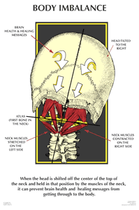
| Home | Evidence | Anatomy | Cervical Spine Biomechanics CERVICAL SPINE BIOMECHANICS Understanding of cervical spine biomechanics is important in understanding the mechanism of any injury to the upper cervical spine. Biomechanics is basically a science, which applies physical and mechanical laws to biological structures like muscles, ligaments, joints and various other structures. Since the human spine is populated with many of these structures in a complex web it is possible for changes in these structures or changes in the position of the skull (occiput or C0) on top of the cervical spine to affect the biomechanical abilities of the cervical spine to hold the head vertical and therefore affect normal movement at that level. Also, because of the close proximity of vital anatomy like cranial nerves, the spinal cord, the brainstem, arteries and other blood vessels it stands to reason that any change in cervical spine biomechanics may well have a detrimental affect on these vital structures and hence affect a person's overall health. We are all accustomed to the disastrous consequences for someone who breaks their neck (cervical spine) or who sustains a dislocation of fracture of cervical vertebrae. Certainly dislocations or fractures in the upper cervical spine are invariably fatal or can be neurologically detrimental. What consequences, symptoms or other problems do people experience that do not exhibit any visible (as viewed on basic X-ray, CT scan or MRI) dislocation or fracture of the cervical spine? Nothing? What happens when a person receives a significant blow to the head, which may or may not result in unconsciousness? Nothing? Just because normal radiographic analysis results in a diagnosis of "Within Normal Limits", does this mean that there has been no damage to cervical spine biomechanics? I suggest not. It's just not plausible that nothing happens to the structures that maintain biomechanical stability. It stands to reason that at the very least ligaments can be stretched briefly and at the other end of the scale stretched beyond their elastic limits or even tear. In these cases it can be the very anatomy of a person, their age and their physical strength that determines whether they are fatally inflicted or just have some minor neck pain. Of course, there are many people in between who have chronic pain and dysfunction for years yet Doctors can find nothing wrong with these people using the tools and methods at their disposal. All things, which are influenced by gravity, are normally stable when the centre of gravity is in synchronisation with the forces and weights affecting them. Thus it is clear that a structure like the human spine with the head sitting atop the cervical spine is mechanically stable when the head is directly over the pelvis. A biomechanically stable spine is characterised by a head sitting vertical to the cervical spine and the eyes, jaw, shoulders and pelvis, which are level with the horizon. There should be neither rotation of the head, shoulders, pelvis nor any anterior or posterior lean of the spine from the cervical spine down to L5. Any deviation from the centre will induce axial loading forces, and alter the weight bearing structures throughout the body. No more is this evident than in the cervical spine. Changes in the biomechanical structures holding the skull on to the atlas vertebra will alter the weight bearing capability of the cervical spine. This resultant change in the centre of gravity can cause postural asymmetry, which represents a mechanical and physiological imbalance of the spine. Injury to ligaments attached to the atlas and skull can result in a complete shift of the skull on the atlas. According to White and Panjabi1 page 283, "the anatomic structures which provide stability for the articulation of the occipital-atlanto-axial articulations are the anterior and posterior atlantooccipital membranes, tectorial membrane, alar ligaments and apical ligaments." I think we can also add some of the sub-occipital ligaments like the rectus capitis posterior minor (RCPMI) and major, obliquus capitis superior and inferior. The RCMPI attaches to the posterior arch of the atlas, to the occiput and via the Myodural Bridge to the dura mater. Form comments from other authors White and Panjabi note that these authors "believe that the occipital-atlantal joint is relatively unstable, at least in children." What I notice most about sick children I have seen is their inability to hold their head up vertical and their tendency to hold their heads in forward posture. I also notice that some young children have very large heads, almost the size of adults (which weight about 4 to 5kg) yet their necks seem so frail as to seem incapable of holding the head upright on the neck. Are these necks unstable as defined in the literature? White and Panjabi provide a table of criteria for instability of the C0-C1-C2 complex. Page 285, Table 5-3.
Case: One particular boy who was suffering from headaches for months along with watery eyes, neck pain and restricted range of motion (ROM) of his cervical spine, exhibited forward head posture. Following an upper cervical chiropractic adjustment to his atlas his normal posture was restored, his headaches disappeared, his eyes stopped watering and his ROM returned to normal, and that was all within hours! He has the biggest head I have ever seen on a child with a really tiny neck and he was only 7 years of age. His GP was busily having him CT-scanned looking for a tumour. The boy's mother was over the moon and the boy returned to the sport he loved. The GP was sceptical and figured the boy was going to grow out of the headaches. In other words it was just a coincidence! I guess this is an anecdotal case, although I witnessed it so it is not anecdotal to me. So be it! Send me more sick kids so I see an anecdotal, coincidental procedure performed on them!
1 Clinical Biomechanics of the Spine- Second Edition
1990; White and Panjabi
|
 The
question I think becomes, "If the C0-C1-C2 complex is studied on
a particular person and the criteria for 'instability' are
not met, does this mean that the person is considered to be 'within
normal limits'" and therefore there is nothing wrong with
them? Maybe even smaller deviations than the limits in the above table
have negative affects on a person's health, which manifest themselves
as symptoms I have described in my own case or as suffered by the boy
in the following case.