
 |
 |
| Home | Evidence | Anatomy |
Grostic Measurment & Analysis GROSTIC MEASURMENT
& ANALYSIS The Origins Of The Grostic Procedure ABOUT THE AUTHOR HISTORY Dr. John F. Grostic was one of the members of this organization. He, along with other chiropractors, would present research and ideas at the annual meeting of the Council. This annual meeting evolved into the Pre-Iyceum program where it continued to be the forum at which new ideas could be presented. At these forums and in the "Bulletin" published monthly by the Council, Dr. Grostic presented much of his research work. Since much of the material was being presented as it was being developed, the continuity in the presentations was lacking. Because of this, several chiropractors requested that Dr. Grostic assemble his research into a "package" that could then be presented to them at one time. In 1946, Dr. Grostic presented the first seminar of the research work to a group of 14 doctors. At the present time, the Grostic Procedure is being taught at Palmer College of Chiropractic as an elective course for senior students and it also is being offered to practicing chiropractors through Palmer College Postgraduate Education Seminars. The Grostic Procedure is primarily a measurement system. The x-ray analysis is the real core of the procedure and is the one area that has remained constant over the last 30 years. During that time, the adjusting methods have changed several times in an effort to improve the effectiveness of the procedure. Since 1946, the adjustment has changed from a Palmer Toggle to what may still resemble a "Toggle," but which is now a much shorter and lighter thrust. The contact point, the pisiform, usually travels less than one-fourth inch during the thrust. The result of this shortened thrust has been twofold. First, discomfort for the patient has for the most part been eliminated. Second, and more important, the atlas misalignment can be reduced more consistently and predictably. The Grostic Procedure originated as a means of precisely measuring the misalignments of the atlas and axis and this is still its prime function. It provides a means of evaluating various adjusting procedures. Because of this evaluating ability, new adjusting methods are continually being tested. When a particular change is proven to reduce misalignments better, it is incorporated into the adjusting aspect of the Grostic Procedure. The procedure begins with the most basic chiropractic philosophy -- that the human body has an inborn intelligence that controls function, growth and repair, and that this inborn intelligence must maintain a balanced and stable relationship between the body and its internal as well as its external environment. It is the function of the nervous system to maintain this homeostasis. To maintain this critical balance effectively, the nervous system must be continually informed not only of any changes in the external environment, but also of the current state of all internal systems. To accomplish this task, large amounts of data must be continually transmitted over the nerves to the brain where this information is integrated and acted upon. If a response is required, it is transmitted over nerves to the appropriate structure, organ or cells. Any interference with the transmission of data or of the response can upset the delicate balance of homeostasis. The Grostic Procedure attempts to correct or reduce misalignments that have produced subluxations and their resulting nerve interference. The reduction of these subluxations allows the data and the response to again travel between the body and the central nervous system re-establishing homeostasis. This procedure provides a precise means of evaluating a misalignment of the upper cervical region of the spine. The misalignment can be a lateral or rotational misalignment of the atlas with respect to the skull or with respect to the axis or any combination of these misalignments. Of course, a misalignment does not always produce a subluxation and the presence of a subluxation must be determined by clinical evaluation of the patients. But, if misalignments are said to produce subluxations; one should be able to cite the mechanism by which this occurs. Based on current knowledge, it would appear that there are at least four major mechanisms by which a misalignment of the upper cervical area of the spine can produce nerve interference and possible nerve dysfunction:
The Grostic Procedure did not dictate the "normal
position" of the atlas. It instead provided a system of measurement
that made possible the locating of that position of the atlas that resulted
in the removal of abnormal clinical findings for the greatest period of
time. This procedure no more dictates the "normal position"
of atlas than physiology texts dictate the normal oral temperature to
be 98.6 degrees. This system of measuring atlas laterality is similar to the other methods that have been used over the years. Except, where other methods have used the tops of the ocular orbits, the tips of the mastoids, the jugular processes, or the inferior tips of the condyles; the Grostic Procedure utilizes the skull itself. This method uses a statistical averaging of the many points that make up the side of the skull rather than choosing any one point, such as the tip of the condyle, as being representative of the entire skull. The Grostic Procedure also assumes that the atlas line, drawn through the attachment points, should be very close to perpendicular to a line drawn through the center of the lower cervical spine. The lower cervical spine line is drawn from a point bisecting the distance between the lateral margins of the left and right zygapophyseal articular surfaces of the sixth or seventh cervical vertebra through a point midway between the center of the odontoid and the superior tip of the spinous process. The fourth major assumption is that the odontoid* and spinous process of C-2 should also be positioned at the center of the atlas as viewed on the nasium view. *In about 20% of the cases, the odontoid is laterally displaced. In these cases, the center of the odontoid is not in the center of the axis. It is necessary to use the actual center of the axis when this condition is present. The center is found by bisecting the superior surface of axis. REFERENCES
|
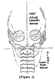
 Several
assumptions about the skull and cervical spine are made by the Procedure.
The first assumption is that the skull on a nasium x-ray view is an incomplete
elipsoid. This elipsoid has a major and minor axis and the major axis
will be referred to as the vertical-central-skull-line. This line can
be determined by using a skull measuring device, the cephlocentroscope,
or by various mathematical methods of fitting an elipse through man points.
It is assumed that this vertical-central-skull-line should be very close
to vertical when the patients are free of subluxation and in the upright
position. (Figure 1)
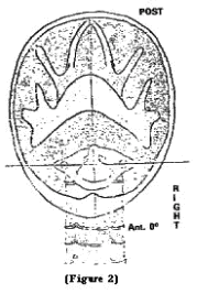
 The
Grostic Procedure makes a third assumption that the atlas viewed on the
vertex or base-posterior x-ray view should be positioned so that a line
drawn through the centers of the foramen transversarium will be nearly
perpendicular to a line longitudinally bisecting the skull. (Figure
2)