Home | Evidence | Anatomy |
Upper Cervical Animations
UPPER CERVICAL ANIMATIONS

These animations and the following ones, which have been downloaded from
the now defunct Life University Research site, are valuable in explaining
the mechanisms of injury and of the corrective adjustment to the upper
cervical spine. Hopefully, I can give you my layperson’s interpretation
to make it easy for you to understand what you are viewing.
 This
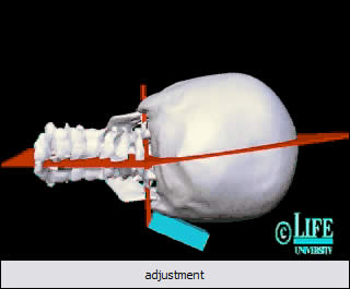
animation is of an upper cervical adjustment. This adjustment
shows how the head and neck are re-aligned after an upper cervical
(atlas) adjustment is performed by an upper cervical chiropractor.
The headpiece serves to allow the head to drop and for the relationship
between the skull and atlas to be restored. This type of dropping
head piece is typically used in toggle recoil adjustments to the
atlas. This
animation is of an upper cervical adjustment. This adjustment
shows how the head and neck are re-aligned after an upper cervical
(atlas) adjustment is performed by an upper cervical chiropractor.
The headpiece serves to allow the head to drop and for the relationship
between the skull and atlas to be restored. This type of dropping
head piece is typically used in toggle recoil adjustments to the
atlas.
 This
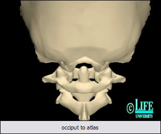
animation emphasizes what I consider to be quite a common injury
to the upper cervical spine. In playing the movie you will note
the how the skull, when receiving a force to one side, glides up
the occipital condyles on the atlas (C1) vertebra and ‘jams’
the atlas in a subluxated position. Use the slider in the Media
Player to view this mechanism of injury. Now envisage the tension,
which must be placed on the spinal cord and lower brainstem, as
well as on critical structures, neurological and vascular exiting
the skull. This
animation emphasizes what I consider to be quite a common injury
to the upper cervical spine. In playing the movie you will note
the how the skull, when receiving a force to one side, glides up
the occipital condyles on the atlas (C1) vertebra and ‘jams’
the atlas in a subluxated position. Use the slider in the Media
Player to view this mechanism of injury. Now envisage the tension,
which must be placed on the spinal cord and lower brainstem, as
well as on critical structures, neurological and vascular exiting
the skull.
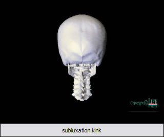 This
animation is known as the ‘Grostic Kink Subluxation’
and shows the effects of an upper cervical subluxation on head and
neck alignment. Use the Media Player slider to view the animation
frame by frame. Note the pronounced misalignment and angle of the
neck in relation to the skull. This is one of the hallmarks of someone
who has sustained an atlas subluxation and if you are alert you
will spot people with this problem, daily. This
animation is known as the ‘Grostic Kink Subluxation’
and shows the effects of an upper cervical subluxation on head and
neck alignment. Use the Media Player slider to view the animation
frame by frame. Note the pronounced misalignment and angle of the
neck in relation to the skull. This is one of the hallmarks of someone
who has sustained an atlas subluxation and if you are alert you
will spot people with this problem, daily.
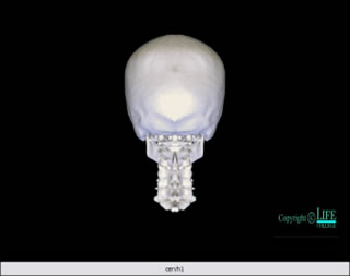 This
is an animation that gives a 360° panoramic view of the skull
atop the cervical spine. If you stop the animation halfway through,
you can view the atlas in the open mouth position in its position
at the back of the throat. This
is an animation that gives a 360° panoramic view of the skull
atop the cervical spine. If you stop the animation halfway through,
you can view the atlas in the open mouth position in its position
at the back of the throat.
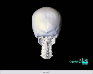 This
is an animation of the cervical spine in rotation. Note how the
vertebrae move as a ‘global’ unit with the atlas exhibiting
the greatest amount of rotation, and the base of the cervical
spine (C7) rotating the least. Subluxations in the upper cervical
region can and do interfere with the biomechanical integrity of
the whole cervical spine and affect its ability to work as a complete
and contiguous unit. This
is an animation of the cervical spine in rotation. Note how the
vertebrae move as a ‘global’ unit with the atlas exhibiting
the greatest amount of rotation, and the base of the cervical
spine (C7) rotating the least. Subluxations in the upper cervical
region can and do interfere with the biomechanical integrity of
the whole cervical spine and affect its ability to work as a complete
and contiguous unit.
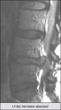 This
is a file from the Society of Orthospinology website www.orthospinology.org and shows the amazing reabsorption of the L4 disc which had herniated
into the spinal canal. The upper cervical chiropractic technique
employed was ‘Atlas Orthogonal’ described elsewhere
on my website, and the period of care and adjustment for this
person was 8 months. This morph of sagittal MRI taken over that
period clearly serves to highlight how adjustments made to the
upper cervical spine (atlas) and re-positioning of the skull so
that it is placed directly over the spinal column and pelvis below
it, results in complete biomechanical changes, resulting in re-alignment
all the way to the base of the spine. This
is a file from the Society of Orthospinology website www.orthospinology.org and shows the amazing reabsorption of the L4 disc which had herniated
into the spinal canal. The upper cervical chiropractic technique
employed was ‘Atlas Orthogonal’ described elsewhere
on my website, and the period of care and adjustment for this
person was 8 months. This morph of sagittal MRI taken over that
period clearly serves to highlight how adjustments made to the
upper cervical spine (atlas) and re-positioning of the skull so
that it is placed directly over the spinal column and pelvis below
it, results in complete biomechanical changes, resulting in re-alignment
all the way to the base of the spine.
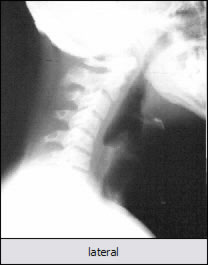 Again,
this is a file from the Society of Orthospinology website www.orthospinology.org and shows the change in cervical lordosis which is achieved when
the skull is re-positioned, by atlas adjustment, to its rightful
position. This position requires having the centre of gravity
over the C7 vertebra. Again,
this is a file from the Society of Orthospinology website www.orthospinology.org and shows the change in cervical lordosis which is achieved when
the skull is re-positioned, by atlas adjustment, to its rightful
position. This position requires having the centre of gravity
over the C7 vertebra.
|

